Teeth bleaching Light-Scam? Depression/Suicide among Dentists
UV Light-Enhanced Tooth Bleaching, A Scam ?
Photochem. Photobiol. Sci., Feb, 2009
The use of optical radiation in the so-called light-assisted tooth bleaching procedures has been suggested to enhance the oxidizing effect of the bleaching agent, hydrogen peroxide. Documentation is scarce on the potential adverse effects of bleaching products and on optical exposure risks to eyes and skin. The efficacy of seven bleaching products with or without simultaneous use of seven different bleaching lamps was investigated using extracted human teeth. The bleaching effect was determined immediately after treatment and one week later. Tooth surfaces were examined for adverse alterations after bleaching using a scanning electron microscope. Source characteristics of eight lamps intended for tooth bleaching were determined. International guidelines on optical radiation were used to assess eye and skin exposure hazards due to UV and visible light emission from the lamps.
Inspection of teeth one week after bleaching showed no difference in efficacy between teeth bleached with or without irradiation for any of the products. Scratches, probably from the cleaning procedure were frequently seen on bleached enamel irrespective of irradiation. Maximum permissible exposure time (tmax) and threshold limit values were exceeded for about half the bleaching lamps investigated. One lamp exceeded tmax even for reflected blue light within the treatment time. This lamp also exceeded tmax values for UV exposure. The lamps were classified as low risk and as borderline to moderate risk according to a relevant lamp standard. Read Full paper
Dentists Facing Depression And Suicide
An article published in the Journal of the Canadian Dental Association claims that many dentists are at risk of suffering from a chronic mood disorder known as dysthymia. It's a condition the Université de Montréal Department of Dentistry is fighting - preventively.
Dysthymia is characterized by loss of appetite, low levels of energy, desperation, excessive anger, social withdrawal and working long hours to compensate for declining performance, troubles in concentration, guilt and suicidal thoughts.
A 2005 study published in the Journal of the American Dental Association claims that 10 percent of the 560 dentists surveyed suffer from this condition. However, only 15 percent of them are followed by a doctor and receive treatment. More Here
Odontogenic infections as the source of acute maxillary sinusitis
Laryngoscope, Feb, 2009
Objective: To identify radiographic features of odontogenic acute maxillary sinusitis and to determine the frequency of a causative dental infection in patients with radiographic evidence of maxillary sinus fluid.
Study Design: Retrospective review of 101 sinus computed tomography scans with unilateral or bilateral maxillary sinus fluid.
Methods: Each maxillary sinus was graded for extent of fluid, degree of mucosal thickening, and presence of dental pathology. Univariate chi-square analysis was used to identify potential radiologic and demographic features predictive of sinus fluid. Multivariate logistic regression was then used to determine which features were independently predictive.
Results:124 of the 202 maxillary sinuses (61%) had sinus fluid. Univariate analysis excluded age, gender, and prior surgery as predictive features. The multivariate analysis included the radiographic features of oroantral fistula, periapical abscess, periodontal disease, projecting tooth root, and dental caries. Of these, only oroantral fistula and the combination of periodontal disease with either a projecting tooth root or periapical abscess were identified as significant sources of maxillary sinusitis. In sinuses that were <1/3>2/3 opacified by fluid, 79% had an identifiable dental source. Mucosal thickening demonstrated a similar relationship with dental sources, so that sinuses having both >2/3 fluid opacification and moderate mucosal thickening were 86% likely to have an identifiable dental source.
Conclusions:Odontogenic infections are often the source of acute maxillary sinusitis, especially if the radiographic findings of sinusitis are severe.
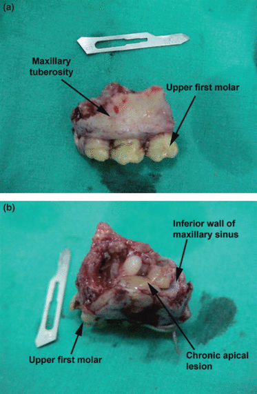
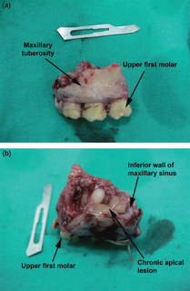 Fracture of the maxillary tuberosity During Extraction
Dental Traumatology Jan 2009
Abstract – Fracture of the maxillary tuberosity sometimes can happen when pneumatization of the maxillary sinus extends between the roots of upper molars. Some factors may lead to this complication including prominent or curved roots, chronic periapical infection, hypercementosis, root ankylosis and
tooth fusion. This paper reports a case with fracture of the maxillary tuberosity following extraction of an upper first molar in general dental practice.
Prevention from any complication during extractions of maxillary molars with large antral enlargement is possible with careful preoperative examination and accurate surgical planning. The general dentist should be prepared to refer such cases to an oral surgeon when facing difficulties like the presented case.
Full Paper
Reattachment of Dehydrated dental fragment using two techniques-Dental Traumatology Jan 2009
Abstract – The reattachment of dental fragments is a conservative treatment and should be considered in the restoration of anterior tooth fractures. This study compared the fracture strength of dehydrated and rehydrated tooth fragments submitted to two different bonding techniques.
Materials and Methods: Sixty human central and lateral mandibular incisors were divided into six groups and sectioned 3 mm from the incisal edge, using a diamond disk. Two reattachment
techniques were applied: (a) bonding, using the Single Bond adhesive system and FiltekZ250 composite resin, followed by placement of a chamfer on the fracture line that was filled with composite resin (Groups 1, 3 and 5); and (b) use of the same bonding technique after dentin removal from the tooth fragment (Groups 2, 4 and 6). The following hydration treatments were applied to the fragments before bonding: (a) 48-h hydration (Groups 1 and 2); (b) 48-h dehydration (Groups 3 and 4); (c) 48-h dehydration followed by rehydration 30 min before
bonding (Groups 5 and 6). The reattached teeth were mounted in acrylic resin cylinders and stored in distilled water for 24 h. The specimens were fractured at a speed of 1 mm min)1
in a universal testing machine.
Results: The following mean fracture strengths (kgf) were recorded: (G1) 12.9 ± 0.6; (G2) 18.8 ± 4.8; (G3) 7.3 ± 1.5; (G4) 15.2 ± 2.4; (G5) 13.4 ± 2.2; and (G6) 17.1 ± 3.2. Analyses using two-way anova and the Tukey test (P < 0.01) revealed significant differences between the restorative techniques and the hydration treatments. Conclusions: The bonding technique that incorporated dentin removal from the fragment before bonding showed greater fracture strength across all groups. Fragment dehydration for 48 h caused a reduction in fracture
strength, which was recovered by a 30-min rehydration. Full paper
Periodontal Treatment Not Found To Reduce Preterm Birth Risk
The study, involving researchers from Duke University Medical Center and the University of North Carolina at Chapel Hill, is one of the largest randomized trials to date to look at the link between the two conditions.
Previous research had suggested that gum disease was associated with very preterm deliveries (defined as less than 32 weeks gestation). That led insurance policies and healthcare providers to recommend scaling and root planing, sometimes referred to as "deep cleaning," in pregnant women. It was thought that such care had the potential to reduce preterm delivery risk.
These new findings, based on a randomized trial of 1,800 pregnant women with periodontal disease, indicate that routine gum treatments do not reduce the risk of early delivery.
Despite the findings, Murtha said much remains unknown about the relationship between the two conditions. "Periodontal disease and poor pregnancy outcomes travel together, but we don't know why." More here
Government Dental College Celebrates Golden Jublee
THIRUVANANTHAPURAM: A year-long programme has been planned to celebrate the golden jubilee of the Government Dental College, Thiruvananthapuram. The function will be inaugurated by Health Minister P.K. Sreemathi on February 8. Different programmes have been charted out for the year, including a mid-year meeting and a get-together of the students present and past; especially those working abroad. This has been planned on July 11 and 12. The valedictory function will be held on December 20. For personal info/enquiry you can call +919633301959 More Here
Photochem. Photobiol. Sci., Feb, 2009
The use of optical radiation in the so-called light-assisted tooth bleaching procedures has been suggested to enhance the oxidizing effect of the bleaching agent, hydrogen peroxide. Documentation is scarce on the potential adverse effects of bleaching products and on optical exposure risks to eyes and skin. The efficacy of seven bleaching products with or without simultaneous use of seven different bleaching lamps was investigated using extracted human teeth. The bleaching effect was determined immediately after treatment and one week later. Tooth surfaces were examined for adverse alterations after bleaching using a scanning electron microscope. Source characteristics of eight lamps intended for tooth bleaching were determined. International guidelines on optical radiation were used to assess eye and skin exposure hazards due to UV and visible light emission from the lamps.
Inspection of teeth one week after bleaching showed no difference in efficacy between teeth bleached with or without irradiation for any of the products. Scratches, probably from the cleaning procedure were frequently seen on bleached enamel irrespective of irradiation. Maximum permissible exposure time (tmax) and threshold limit values were exceeded for about half the bleaching lamps investigated. One lamp exceeded tmax even for reflected blue light within the treatment time. This lamp also exceeded tmax values for UV exposure. The lamps were classified as low risk and as borderline to moderate risk according to a relevant lamp standard. Read Full paper
Dentists Facing Depression And Suicide
An article published in the Journal of the Canadian Dental Association claims that many dentists are at risk of suffering from a chronic mood disorder known as dysthymia. It's a condition the Université de Montréal Department of Dentistry is fighting - preventively.
Dysthymia is characterized by loss of appetite, low levels of energy, desperation, excessive anger, social withdrawal and working long hours to compensate for declining performance, troubles in concentration, guilt and suicidal thoughts.
A 2005 study published in the Journal of the American Dental Association claims that 10 percent of the 560 dentists surveyed suffer from this condition. However, only 15 percent of them are followed by a doctor and receive treatment. More Here
Odontogenic infections as the source of acute maxillary sinusitis
Laryngoscope, Feb, 2009
Objective: To identify radiographic features of odontogenic acute maxillary sinusitis and to determine the frequency of a causative dental infection in patients with radiographic evidence of maxillary sinus fluid.
Study Design: Retrospective review of 101 sinus computed tomography scans with unilateral or bilateral maxillary sinus fluid.
Methods: Each maxillary sinus was graded for extent of fluid, degree of mucosal thickening, and presence of dental pathology. Univariate chi-square analysis was used to identify potential radiologic and demographic features predictive of sinus fluid. Multivariate logistic regression was then used to determine which features were independently predictive.
Results:124 of the 202 maxillary sinuses (61%) had sinus fluid. Univariate analysis excluded age, gender, and prior surgery as predictive features. The multivariate analysis included the radiographic features of oroantral fistula, periapical abscess, periodontal disease, projecting tooth root, and dental caries. Of these, only oroantral fistula and the combination of periodontal disease with either a projecting tooth root or periapical abscess were identified as significant sources of maxillary sinusitis. In sinuses that were <1/3>2/3 opacified by fluid, 79% had an identifiable dental source. Mucosal thickening demonstrated a similar relationship with dental sources, so that sinuses having both >2/3 fluid opacification and moderate mucosal thickening were 86% likely to have an identifiable dental source.
Conclusions:Odontogenic infections are often the source of acute maxillary sinusitis, especially if the radiographic findings of sinusitis are severe.
 Fracture of the maxillary tuberosity During Extraction
Dental Traumatology Jan 2009
Abstract – Fracture of the maxillary tuberosity sometimes can happen when pneumatization of the maxillary sinus extends between the roots of upper molars. Some factors may lead to this complication including prominent or curved roots, chronic periapical infection, hypercementosis, root ankylosis and
tooth fusion. This paper reports a case with fracture of the maxillary tuberosity following extraction of an upper first molar in general dental practice.
Prevention from any complication during extractions of maxillary molars with large antral enlargement is possible with careful preoperative examination and accurate surgical planning. The general dentist should be prepared to refer such cases to an oral surgeon when facing difficulties like the presented case.
Full Paper
Reattachment of Dehydrated dental fragment using two techniques-Dental Traumatology Jan 2009
Abstract – The reattachment of dental fragments is a conservative treatment and should be considered in the restoration of anterior tooth fractures. This study compared the fracture strength of dehydrated and rehydrated tooth fragments submitted to two different bonding techniques.
Materials and Methods: Sixty human central and lateral mandibular incisors were divided into six groups and sectioned 3 mm from the incisal edge, using a diamond disk. Two reattachment
techniques were applied: (a) bonding, using the Single Bond adhesive system and FiltekZ250 composite resin, followed by placement of a chamfer on the fracture line that was filled with composite resin (Groups 1, 3 and 5); and (b) use of the same bonding technique after dentin removal from the tooth fragment (Groups 2, 4 and 6). The following hydration treatments were applied to the fragments before bonding: (a) 48-h hydration (Groups 1 and 2); (b) 48-h dehydration (Groups 3 and 4); (c) 48-h dehydration followed by rehydration 30 min before
bonding (Groups 5 and 6). The reattached teeth were mounted in acrylic resin cylinders and stored in distilled water for 24 h. The specimens were fractured at a speed of 1 mm min)1
in a universal testing machine.
Results: The following mean fracture strengths (kgf) were recorded: (G1) 12.9 ± 0.6; (G2) 18.8 ± 4.8; (G3) 7.3 ± 1.5; (G4) 15.2 ± 2.4; (G5) 13.4 ± 2.2; and (G6) 17.1 ± 3.2. Analyses using two-way anova and the Tukey test (P < 0.01) revealed significant differences between the restorative techniques and the hydration treatments. Conclusions: The bonding technique that incorporated dentin removal from the fragment before bonding showed greater fracture strength across all groups. Fragment dehydration for 48 h caused a reduction in fracture
strength, which was recovered by a 30-min rehydration. Full paper
Periodontal Treatment Not Found To Reduce Preterm Birth Risk
The study, involving researchers from Duke University Medical Center and the University of North Carolina at Chapel Hill, is one of the largest randomized trials to date to look at the link between the two conditions.
Previous research had suggested that gum disease was associated with very preterm deliveries (defined as less than 32 weeks gestation). That led insurance policies and healthcare providers to recommend scaling and root planing, sometimes referred to as "deep cleaning," in pregnant women. It was thought that such care had the potential to reduce preterm delivery risk.
These new findings, based on a randomized trial of 1,800 pregnant women with periodontal disease, indicate that routine gum treatments do not reduce the risk of early delivery.
Despite the findings, Murtha said much remains unknown about the relationship between the two conditions. "Periodontal disease and poor pregnancy outcomes travel together, but we don't know why." More here
Government Dental College Celebrates Golden Jublee
THIRUVANANTHAPURAM: A year-long programme has been planned to celebrate the golden jubilee of the Government Dental College, Thiruvananthapuram. The function will be inaugurated by Health Minister P.K. Sreemathi on February 8. Different programmes have been charted out for the year, including a mid-year meeting and a get-together of the students present and past; especially those working abroad. This has been planned on July 11 and 12. The valedictory function will be held on December 20. For personal info/enquiry you can call +919633301959 More Here
posted by Healthmantra at
6:49 PM
![]()
![]()

0 Comments:
Post a Comment
<< Home