International Academy For Rotary Endodontics
International Academy for Rotary Endodontics (IARE) was launched on 4th July 2005. World class clinical facility was
established to train dentists in latest in Rotary endodontics. Profile of IARE HERE
This center was named after the great innovator and clinician Dr. E Steve Senia. Steve Senia Rotary Endodontic center is
one of the finest centers in the world to learn Rotary Endodontics.If you want the best, this is the place (our participants say so). Since inception
the center has been helping dentitst/endodontists from world over to perform better and efficient root canal treatment.
This means what you learn here you will be able to practice in your clinic the very next day after the course. ARE YOU A DEDICATED DENTIST ?
Fear grips you when you do endo or during obturation, nagging patients keep coming back with complaint of pain during/after endo. You always dream of achieving successful "single sitting" rotary endo, come to Steve Senia center to learn Endo and your life will never be same again.
Mission: To promote quality Endodontics based on science rather than myths.
Vision: To train Dentists and Endodontists who strive for excellence as their life goal.
Read this page and click on the links on right side of this page to browse other sections of Rotary Endo and we promise that you will experience what you have been missing !!!! Rotary Nirvana.
Want to get certified and get International fellowship ?
Click here Fellowship (FIARE, USA)
Marketing guys enjoy making FOOL of YOU, but if you can see for yourself, in a minute you can understand how complex is canal anatomy, excellent work
in this area has been done by Dr. Marco Versiani, see his outstanding work HERE
Image below is from his work, GREEN areas are BEFORE preparation, RED are after preparation, in this overlap pic GREEN shows the areas
that have NOT been touched by endo instruments, MOST likely that is the case with many your patients also.....thats why they come back with
pain and problems.

Do not believe marketing gimmicks. Here are two ways you can see proof yourself.
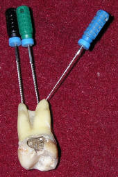
1. When you extract a tooth, see which is the smallest instrument that
can enter from the apex (uninstrumented), often you will see that it is size 30
or larger (see pic below), if you have finished with an F1 or F2 that means that you have not
done any apical cleaning.
You can also cut 1/3 apex of an extracted tooth and see which instrument goes in ?
2.Do RCT on extracted tooth and section apical 1-2
mm and examine under a magnification device. OR do RCT on a tooth which will be extracted (eg. orthodontic etc) and then after extraction examine
tooth under magnification (after sectioning apical 1-2 mm).
This way you will yourself see the TRUE comparison of different systems.
3. What is the basis for apical surgery ? We open and seal the apex, which again is the proof that apical area is the MOST important. BUT when you finish apex 20 or 25 with a tapered system, you will be shocked to see the amount of debris, when you section and examine apical area under magnification.
In a Sept-Oct 2007 issue of General dent. an article by Francis W. Allen points out that in past 20% apical cleaning was achievable with hand instruments, tapered instruments clean only 18% in the apical 1mm, no wonder we spend so much energy on referrals/retreatment, to read the full paper CLICK HERE
Another excellent paper from a leading journal: This explains importance and method of cleaning apical third of root canals for absolute success in single visit endodontics. To Read CLICK HERE
4. Have you heard that during extraction of a tooth if root tip breaks, easiest way is to screw in a headstrom file and root tip easily comes out. And do you know what file size is normally used ? 35 yes that shows the canal size in apical area is larger than 35 (Ref is JADA: STONER 133 (4): p 473, 2002) to read full ref click HERE
People show coronal pictures (scopes) all the time, its surprising why NO ONE shows pics of apical sections. It is a proven fact that apical area is the most CRITICAL, if you do not clean it well your chances for FAILURE are much higher.
ARE you guilty of not doing the best, Give the Very Best to your patient CLICK HEREIntra oral X ray shows only two dimensional view, 3D systems are right now very expensive. So IOPA can not be a criteria to judge quality of RCT and there can be no proper RCT without APEX/ Foramen locator.
Endodontics-Make Right Choice to SucceedPracticing exclusive endodontics for over thirty five years, I have developed a systematic/scientific method for choosing right type instruments and techniques that will give predictable results for root canal treatment. Tips and tricks that you will learn with me are priceless.
The concept of Coronal Flaring came as a major breakthrough in endodontics. Instead of heading directly to the apical area, this approach cleaned up the coronal part, of thousands of micro organisms, thus preventing their entry to the apical part. But an unfortunate thing that has happened is that while so much stress is being given to the coronal enlargement, the apical area has been neglected.
Every Endodontist knows that the apical third of the root canal is the critical area that influences the outcome of root canal treatment. It has to be thoroughly cleaned, enlarged and given a hermetic seal. Can we enlarge this critical area to just #20 or 25 instrument and obturate? How many canals will then have hermetic seal, with the exception of those few cases where the canals were calcified and very narrow to start with?
The role of irrigating solutions ( eg.
Sodium Hypochlorite ) in root canal treatment has never been
clearer. For the irrigant to even reach the apical region, this area
needs to be enlarged to minimum # 30. Increase in file size was shown to be
important in allowing the NaOCl to be an effective antibacterial
irrigant.
Journal of Endodontics. 26(12):751-755, December 2000. Shuping, George B. DDS, MS; Orstavik, Dag DDS, PhD; Sigurdsson, Asgeir DDS, MS; Trope, Martin DMD )
When apical region is not cleaned (particularly in infected cases) if you section and see apex under magnification, you can see debris (pic on left) and such case will fail, needing retreatment/apical surgury.
A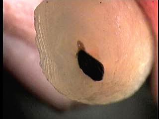 |
B |
There is enough literature to show that uninstrumented apical diameter is large (.3 -.6) but we have been blinded by marketing forces. See this Recent Publication
There are too many concepts promoted without any scientific basis to divert your attention from core issues which are vital for endodontic success. One concept is that lateral canals should be cleaned and obturated and secondly it is better to be apical barbarian then to be pulp lover. Here we bring you two great scientific studies which clear these doubts. CLICK HEREWe have compiled some more evidence to show you where tapered instrument binds and HOW it does not clean the apical AREA. CLICK HERE to See this exciting ENDO TRUTH.
To attend our course SEE course brochure OR click Here to Enroll Tweet