Tuesday, May 27, 2008

Bihar Health Dept Outsources Dental Care

Bihar Health Department Outsources Dental Care

PATNA: The health department has decided to set up dental clinics at all the district (sadar) and sub-divisional hospitals of Bihar under the National Rural Health Mission. These dental clinics would be set up under public-private partnership (PPP).

Under this partnership, while the state government will provide land for the dental clinics, the private agencies, willing to enter the partnership, would have to provide machines and equipment for the clinics. These agencies would also be responsible for maintenance and running of the clinics and in turn they would be allowed to take fee from the patients.

The selection of private agencies, which would be for a period of five years, would be made on the basis of maximum amount offered to the state government in lieu of land provided by it.

Agencies interested in setting up dental clinics would have to submit a proposal of minimum Rs 1.25 lakh for one unit, Rs 3 lakh for units numbering from 2 to 5, Rs 5 lakh for 6 to 10 units, Rs 10 lakh for 11 to 20 units and Rs 50 lakh for running all such units.

Apart from the agencies, individuals having a BDS degree are also eligible for making bids for setting up the dental clinics.

Lets hope Whole of India adopts novel strategies for Dental Care of Indians.

Piercings can cause dental problems

Body piercings, especially to the lips and tongue, can cause serious dental complications, according to a research.
In the study conducted by the University of Tel Aviv on 400 consecutive patients, who were aged 20 years on average, every fourth person with a piercing in the tongue or lips revealed symptoms such as gum bleeding.
Some 13.9 percent had broken teeth or other dental complications, the study found.
Dental professionals were warned of the increasing number of patients with oral piercings and to provide appropriate guidance to patients regarding the health risks.


16 May, 2008- 3M Acquires IMTEC

3M announced today that it has signed a definitive agreement to acquire IMTEC Corp., a manufacturer of dental implants and cone beam computed tomography (CBCT) scanning equipment for dental and medical radiology, Full news HERE

Higher Cancer Risk For Those With Gum Disease

Whether they are smokers or non-smokers, people with gum disease have a higher overall risk of cancer, according to an Article published on May 27, 2008 in The Lancet Oncology.

Gum disease, such as periodontis or gingivitis, is associated with increased concentrations of inflammatory markers in the blood. There is some debate, however, about whether this systemic inflammation, the pathogenic invasion into the blood stream, or the immune response to gum infection could possibly affect cancer risk, overall or at specific sites. MORE HERE


And there is this nice patient education article worth looking at
Look after your teeth and gums and your whole body will start to feel the benefit

Saturday, May 24, 2008

World' s Smallest Laser & Disappearing Roots

World's Smallest Laser

Dental laser industry leader Zap Lasers unveiled a breakthrough in soft-tissue management, the Styla MicroLaser™. The world's first microlaser for soft-tissue applications, the 1.9 ounce Styla combines revolutionary design and proven diode laser technology in a self-contained unit measuring only 6.9 inches long. The handheld Styla has no wires or cables, allowing dental professionals to move freely throughout treatment areas.
Salient Advantages claimed:

-Lightweight Aluminum Body
-Pre-threaded Disposable Tips
-Intelligent Gravity Sensor
-Rechargeable Lithium Ion Batteries
-Wireless Foot Pedal
To see company release of this laser GO HERE

Case Report on Disappearing Roots

Tooth resorption is a perplexing problem for all dental practitioners. The etiologic factors are vague, the diagnoses are educated guesses and, often, the chosen treatment does not prevent the rapid disappearance of the calcified dental tissues. This becomes all the more confusing if the tooth in question is a pulpally involved young permanent tooth. Presented in this report is the case of an upper first young permanent molar that underwent complete root resorption following root canal therapy and obturation.
To Read full Report CLICK HERE

Smoothies Can Damage Your Teeth

Dentists have warned the current popularity of fruit smoothies could lead to widespread tooth damage.

It was claimed last week that the fruit drinks could be twice as healthy as first thought.

But dentists warn that the beneficial effects of boosting consumption of fruit are likely to be far outweighed by damage the drinks cause to teeth.

They warn that the high levels of sugar in the drinks can promote decay without good dental hygiene. To Read the full BBC report CLICK HERE

Oral Health Can Be Affected For Years To Come From Premature Tooth Loss

Two kinds of traumatic tooth loss can occur during childhood: The child can lose a primary (baby) tooth prematurely, or they can lose a permanent tooth. Full report of AGD here Parents are advised to take due care to avoid long term damage.

Thursday, May 15, 2008

Tooth Jewellery and now Tooth Tattoos, Tooth loss leads to Cancer

Tooth Tatoos

You may be familiar with tooth jewellery ? The popular one for quite a while has been the brand known as skyce. These are small crystals looking like diamonds and are fixed on front teeth to give a shine to your smile. If you are not familiar you can click here to learn more. As if this was not enough you now have tooth tattoos.

American tattoo artist Steve Heward, who is also a dental technician, has taken the art of tattooing from the body to the mouth and has made it a huge success. Heward, who has been practising his art since 1979, has also made crowns for a few famous people, including a candidate for the US presidency in 2000.

The tooth artists at Heward Dental Lab can create custom hand-painted artwork and paint celebrities on your tooth. "The work can take from five minutes to an hour. But I usually spend half an hour," said Heward.

And what are the damages? "For the artwork itself, I usually charge about $75 to $500 depending on the subject matter," said Heward.

"My crowns can last a lifetime. And removing the tattoos is also a brief and painless process."

And there are people who love to put pictures of teeth tattoo on their bodies, SEE HERE

Tooth Loss Linked To Esophageal, Head And Neck, And Lung Cancer

Bad dental health-infected Teeth/Gums is a major HEALTH HAZARD, this point has to be driven home to our mass population. We have been bringing relevant publications to your mailbox so that in turn you can pass this info to public. To see them all CLICK HERE

Studying thousands of patients, Japanese researchers have found a strong link between tooth loss and increased risk of three cancers esophageal, head and neck, and lung. They suggest that preservation of teeth may decrease risk of developing these diseases.

In the May issue of Cancer Epidemiology, Biomarkers and Prevention, a journal of the American Association for Cancer Research, scientists from Aichi Cancer Center in Nagoya and Nagoya University Graduate School of Medicine speculate that bacterial infection and inflammation resulting from poor oral care that leads to tooth loss could also be driving development of these cancers. Periodontal disease is known to increase risk for stroke and heart disease.

"Tooth loss is a common consequence of chronic bacterial infection and may, therefore, serve as a surrogate for chronic infection and inflammation, which in turn may be important to the pathogenesis of cancer," said the study's lead author, Akio Hiraki, Ph.D., a researcher at the Aichi Cancer Center.

Researchers measured rates of 14 different cancers and rates of tooth loss in 5,240 cancer patients in Japan, and compared those rates among 10,480 matched cancer-free participants. The researchers specifically found that people with tooth loss were 136 percent more likely to develop esophageal cancer, had a 68 percent increased risk of developing head and neck cancer and a 54 percent greater chance of developing lung cancer. The researchers also found that the rate of cancer increased proportionally to the number of teeth a patient had lost.
Conclusion:
There is a significant positive association between tooth loss and the risk of head and neck, esophageal, and lung cancers after adjustment for potential confounding factors. The findings indicate that preventive efforts aimed at the preservation of teeth may decrease the risk of these cancers.

The use of visual assessment of dental radiographs for identifying women at risk of having osteoporosis: the OSTEODENT project published online on triple O, May 2008

Can you identify risk of osteoporosis from Intra oral radiographs? According to this article there is very high (90%) chance of predicting by seeing the trabecular density in premolar region.

Objective

The objective of this study was to investigate the diagnostic accuracy of visual assessment of the trabecular pattern in intraoral periapical radiographs to identify female subjects at risk of having osteoporosis.
Study design

Six hundred female subjects underwent intraoral periapical radiography of the maxillary and mandibular premolar region. Five observers assessed the trabecular pattern as dense, heterogeneous, or sparse, with the aid of reference images. All patients received a central dual energy x-ray absorptiometry (DXA) examination of the hip and lumbar spine.
Results

With sparse trabecular pattern as indicative of osteoporosis, mean specificity was high (91.6 for the upper jaw and 90.8 for the lower jaw) while the sensitivity was low (28.2 for the upper and lower jaw). The mean intraobserver agreement was comparable for radiographs of the upper and lower jaw (median κw 0.53 and 0.57, respectively).
Conclusion

Visual assessment of the trabecular pattern in intraoral periapical radiographs of premolar regions is a potential method to identify women at risk of having osteoporosis.


Labels: , , , ,

Wednesday, May 14, 2008

Local Anaesthesia Reversal and Gums and New born connection

Local Anaesthesia Reversal 10 May

Drooling, biting your lip and talking like you have marbles in your mouth after a visit to the dentist could soon become a thing of the past.

A drug that reverses the numbing effects of local anesthesia used by dentists was approved yesterday by the Food and Drug Administration for patients as young as 6 years old.

OraVerse, which was developed by private San Diego company Novalar, is the first dental anesthesia reversal agent on the market. It cuts in half the time it takes for Novocaine and other numbing agents to wear off, according to clinical studies.

That amounted to about 85 fewer minutes of numbness during clinical trials. About 40 percent of patients in the trials felt sensation return within an hour of receiving the OraVerse injection.
Full Report Here

Molar Distalisation with Mini implants

This has been proved in number of studies that micro or mini implants in orthodontic patients can cut treatment time upto 40%. In the last post we had posted two studies from AJODO which show how quality of treatment would be better with MIA because of better anchorage management. What are you waiting for ? start using microimplants now, they are easy to use and inexpensive, learn more now.

Here we have posted a pic of molar distalisation done in less than six months, start date is 5 Nov 2007 and second pic is taken on 12 May 2008, if you see facial change you will be happy -to see all the intraoral and facial pictures CLICK HERE.


Periodontal disease and adverse birth outcomes: a study from Pakistan
American Journal of Obstetrics and Gynecology -Volume 198, Issue 5, May 2008

There have been several reports that gingival condition during pregnancy should be maintained well to avoid adverse outcomes. This study once again proves that point. Our goal should be to cater to the needs of each and every pregnant lady and Gynecologists should be educated to refer all such patients for routine maintenance and treatment.

Study Design

This was a prospective cohort study. Enrollment occurred at 20-26 weeks of gestation. A study dentist performed the periodontal examination to assess probing depth, clinical attachment level, gingival index, and plaque index. Outcomes included stillbirth, neonatal death, perinatal death, <32 style="font-weight: bold;">Results


Dental examinations and outcome data were completed for 1152 women: 81% of the women were multiparous, with a mean age of 27 years; 33% of the women had no education. Forty-seven percent of the women had dental caries; 27% of the women had missing teeth, and 91% of the women had had no dental care in the last year. Periodontal disease was common: 76% of the women had ≥3 teeth with a probing depth of ≥3 mm; 87% of the women had ≥4 teeth with a clinical attachment level of ≥3 mm; 56% of the women had ≥4 teeth with a plaque index of 3; and 60% of the women had ≥4 teeth with a gingival index of 3. As the measures of periodontal disease increased from the 1st to 4th quartile, stillbirth and neonatal and perinatal death also increased, with relative risks of approximately 1.3. Early preterm birth increased, but the results were not significant. Late preterm birth and low birthweight were not related to measures of periodontal disease.

Conclusion

Pregnant Pakistani women have high levels of moderate-to-severe dental disease. Stillbirth and neonatal and perinatal deaths increased with the severity of periodontal disease.

Sunday, May 11, 2008

Fraudulent Use of Digital Radiography, and Microimplants


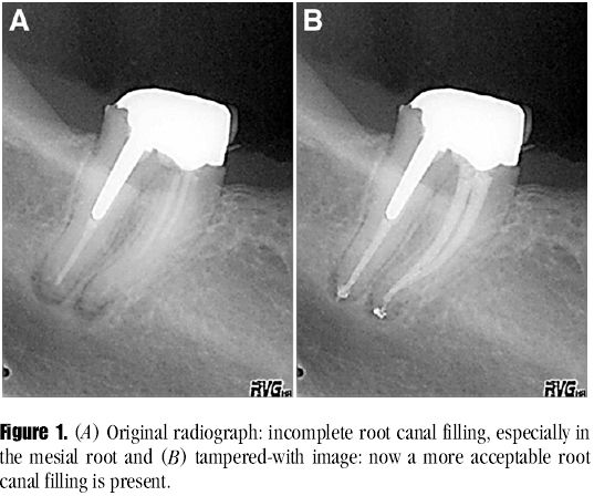
Fraudulent Use of Digital Radiography: Methods To Detect and Protect Digital Radiographs

This very important article is published in Journal of Endodontics, May 2008 issue. As the use of digital radiography is becoming commonplace in dental clinics, it is better to be educated on these issues rather then being fooled by some computer savvy dentists who may show you great cases in their jazzy presentations.

To improve vision and diagnosis, dental x-ray software allows image enhancement (eg, adjusting color, density, sharpness, brightness, or contrast). Exporting digital radiographs to a file format compatible with commercial graphic software increases chances that information can be altered, added, or removed in an unethical manner.
Dental radiographs are easily duplicated, stored, or distributed in digital format. It is difficult to guarantee the authenticity of digital images, which is especially important in insurance or juridic cases. Image-enhancement features applied to digital radiographs allow mishandling or potential abuse. This has been illustrated by several recently published studies. A standard authentication procedure for digital radiographs is needed. A number of manipulated radiographic images are presented to show concerns about security, reliability, and the potential for fraud. Antitampering techniques and methods of detecting manipulations in digital medical images are discussed. See the pic above, one example of manipulation. To request full paper email to icdr@hotmail.com

Evaluation of the risk of infection through exposure to aerosols and spatters in dentistry

Aerosols in clinic can be dangerous ? American Journal of Infection Control May 2008 issue.

Many dental procedures produce extensive aerosols and splatters that are routinely contaminated with microorganisms.
Methods
Air containing blood-bearing aerosols and surfaces contaminated by sedimenting blood particulate was sampled in 5 different dental cubicles. To assess contamination by blood particulate, the concentration of hemoglobin (Hb) in the air and on the sedimentation surfaces was determined.
Results
The mean concentration of Hb in the air aspirated in the 5 cubicles was 0.14 ± 0.23 μg/m3, corresponding to a blood volume of 8.7 × 10⁻⁴ μL/m3. Similarly, the mean concentration of blood particulate sedimented on surfaces was calculated and found to be 1.56 μL/m2. In 80% of the cubicles monitored, 100% positivity to the Hb determination test was recorded in all of the surface samples.
Conclusions
The results obtained revealed contamination of both air and surfaces by blood particulate. Moreover, with the exception of those obtained in 1 cubicle, all of the samples of sedimenting particulate analyzed were positive for the presence of Hb.


Microimplants are here to stay, have you started using ? Come Try now DO IT NOW

Comparison of the intrusion effects on the maxillary incisors between implant anchorage and J-hook headgear --AJODO May 2008, Toru Deguchi etal

Introduction: Recently, miniscrews have been used to provide anchorage during orthodontic treatment, especially for incisor intrusion. Miniscrews during incisor intrusion are commonly used in implant orthodontics. Traditionally, effective incisor intrusion has been accomplished with J-hook headgear. In this study, we compared the effect of incisor intrusion, force vector, and amount of root resorption between implant orthodontics and J-hook headgear. Methods: Lateral cephalometric radiographs from 8 patients in the implant group and 10 patients in the J-hook headgear group were analyzed for incisor retraction. The estimated force vector was analyzed in the horizontal and vertical directions in both groups. Root resorption was also measured on periapical radiographs.
Results:
In the implant group, significant reductions in overjet, overbite, maxillary incisor to palatal plane, and maxillary incisor to upper lip were observed after intrusion of the incisors. In the J-hook headgear group, significant reductions in overjet, overbite, maxillary incisor to upper lip, and maxillary incisor to SN plane were observed after intrusion of the incisors. There were significantly greater reductions in overbite, maxillary incisor to palatal plane, and maxillary incisor to upper lip in the implant group than in the J-hook headgear group. Estimated force analysis resulted in significantly more force in the vertical direction and less in the horizontal direction in the implant group. Furthermore, significantly less root resorption was observed in the implant group compared with the J-hook headgear group. Conclusions: The maxillary incisors were effectively intruded by using miniscrews as orthodontic anchorage without patient cooperation. The amount of root resorption was not affected by activating the ligature wire from the miniscrew during incisor intrusion.


Evaluation of orthodontic mini-implant anchorage in premolar extraction therapy in adolescents
AJODO, May 2008
Introduction: The purpose of this study was to determine the success rate, positional stability, and patient evaluation of orthodontic mini-implants (OMIs). Methods: Thirteen patients (8 girls, 5 boys; average age, 14 years 10 months) were treated with 82 OMIs measuring 1.6 mm in diameter and 6 mm in length placed in the buccal alveoli (1 unloaded OMI and 1 loaded OMI per quadrant). The right or left side of each arch was randomly selected for immediate loading with up to 250 g of direct force; the contralateral side was loaded 3 to 5 weeks later. Serial impressions, clinical observations, and orthodontic maintenance were performed until adequate space closure was achieved.
Results: The overall OMI success rate was 70.73%. As calculated with a mixed-model analysis, there was no statistically significant difference between the success rates of immediately loaded OMIs (80.0%) and delayed loaded OMIs (80.95%). The combined success rate for loaded OMIs (80.49%) was significantly higher than that of unloaded OMIs (60.98%). Patients' motivation for OMI treatment was primarily the desire to avoid headgear. Using a 100-mm visual analog scale, the patients indicated average scores of 54.77 for the amount of pain during OMI placement and 27.10 for the amount of pain during OMI removal.
Conclusions:
OMIs are a predictable, effective, and well-tolerated anchorage source for adolescents. Neither the timing of force application nor the force itself precipitated failure of the OMIs. Orthodontic forces can be applied immediately to OMIs. Various anatomic and behavioral conditions unique to adolescents and a clinical learning curve can affect the success rate of OMIs.


Tuesday, May 6, 2008

Latest news in Dentistry

Treatment and resolution of a mental nerve paresthesia

NOTE: This case is a classic example of inadequate apical cleaning, which can be achieved only by non tapered endo instruments

Several such highly infected cases and with sinuses heal beautifully without the need for apical resection if you use the above mentioned instruments, try it today.

The purpose of the present case report (triple O May 2008 online issue) is to present the treatment and resolution of a mental nerve paresthesia (MNP) stemming from apical pathosis of a mandibular canine tooth and the follow-up of 3 years.

A 28-year-old man was referred to the Selçuk University Faculty of Dentistry Oral-Maxillofacial Surgery Clinic by his dentist in March 2004. The patient had a complaint of slight swelling and numbness of the left skin mucosa of the lower lip and left chin. He also stated that certain swelling disappeared after therapy subsequent to antibiotic and analgesic prescription of the dentist. Even though the certain swelling almost disappeared, the numbness of the lower lip and the pain of the adjacent teeth was refractory.
Radiographic examination revealed a unilocular lesion surrounding the apical portion of the left lower canine tooth. RCT was carried out 3 weeks back. Patient was given antibiotic which did not help and so it was decided to operate on the tooth with apical resection.

At the follow-up appointment, 10 weeks later after first visit, the patients was free of pain and there was a complete resolution of paresthesia and related tooth was asymptomatic. At the recall visit after 3 years from the initial treatment, the patient was still in comfort without any previous symptoms. Panoramic and periapical view of the area showed that peripical radiolucency had been diappeared and the tooth was restored with composite filling without problem.

Message:
ENDO is easy, but good endo is not easy

Botox ? Is it Safe ?

We had brought you an update about the use of Botox in dentistry
Now a recent animal study has shown that botox can move from injection site to brain.
Scientists injected rats' whisker muscles with botulism toxin. Tests of the rodents' brain tissue found that botulism had been transported to the brain stems, the researchers said in the Journal of Neuroscience published April 2. 2008.
Botulism neurotoxin can disrupt nerve cells' ability to communicate and may change spinal cord circuitry, the authors wrote in the study. Full report here

Eruption rates in Diabetes Mellitus Children

It is published in May 2008 issue of Pediatrics (American Journal)
OBJECTIVE. The objective of this study was to evaluate tooth eruption in 6- to 14-year-old children with diabetes mellitus.

METHODS. Tooth eruption status was assessed for 270 children with diabetes and 320 control children without diabetes. Data on important diabetes-related variables were collected. Analyses were performed using logistic regression models.

RESULTS. Children with diabetes exhibited accelerated tooth eruption in the late mixed dentition period (10–14 years of age) compared to healthy children. For both case patients and control subjects the odds of a tooth being in an advanced eruptive stage were significantly higher among girls than boys. There was also a trend associating gingival inflammation with expedited tooth eruption in both groups. No association was found between the odds of a tooth being in an advanced stage of eruption and hemoglobin A1c or duration of diabetes. Patients with higher body mass index percentile demonstrated statistically higher odds for accelerated tooth eruption, but the association was not clinically significant.

CONCLUSIONS. Children with diabetes exhibit accelerated tooth eruption. Future studies need to ascertain the role of such aberrations in dental development and complications such as malocclusion, impaired oral hygiene, and periodontal disease. The standards of care for children with diabetes should include screening and referral programs aimed at oral health promotion and disease prevention.


Sunday, May 4, 2008

Endodontic Courses and Updates

Market survey has shown that in Indian practices which are on TOP have almost 40% contribution from endo work. And practices that loose are those who avoid endo or do it by giving multiple appointments and take several visits to complete obturation.

One of the main reasons is using only hand instruments or stiff tapered instruments. Come join the revolution with the NON tapered rotary instruments and you will be happy WE guarantee. Come and attend Great line up of endo courses to suit your needs, which are SURE to transform your life. 10-11 May at Bangalore, 19 May at Goa, 25-30 May at Belgaum and 10 June-9 July at Belgaum. 25-30 August, Portland, USA For details email or call, icdr@hotmail.com - 94481 91202

Which is the best analgesic in Endodontic treatment ?

This paper tiltled "Gender differences in Analgesia for Endodontic pain" is published in May 2008 issue of Journal of Endodontics.

Forty-three patients were administered ibuprofen 600 mg, placebo, or pentazocine 50 mg/0.5 mg naloxone in a randomized, double-blinded manner. Beginning immediately after endodontic treatment, patients took the assigned medication every 6 hours for 24 hours and recorded their degree of discomfort on a 100-mm visual analog scale. Statistical analysis of the data showed that ibuprofen 600 mg provided statistically significantly greater analgesia than placebo at 6 and 12 hours (P = 0.0014 and 0.0024), and pentazocine/naloxone provided statistically significantly greater analgesia than placebo at 12 hours (P = 0.0084). Sex-dependent differences were noted within the pentazocine/naloxone group, which showed significantly greater analgesia in females compared with males (P = 0.007).

Dentists adopt unconventional treatment options.

But Rothchild's is a dentist in USA and is member of ADA, his practice is based on a holistic philosophy that says the health of your mouth can affect your entire body.
But mounting research supports what holistic dentists have long believed: Although the mouth may not be the window to your soul, it can reveal potential medical problems in other parts of the body, including the heart, lungs and brain.

And as the connection grows stronger, dentists who focus on treating the whole body as well as the teeth and gums—with prevention, education and nutrition playing a central role—may one day assume the role of a "primary care doctor."

This dentist uses acupuncture, osteopathy and Reiki etc. Read full report here


How to improve your composite restorations

One major cause of failure of composite restorations is micro leakage, can we reduce it by modifying the cavities ? Recent article published in Am Journal of Ped dentistry compared the effectiveness of self-etch and total-etch adhesive systems in bonding to the beveled and nonbeveled margins of primary and permanent teeth.
Methods: This in vitro, factorial-designed study allowed evaluation of 3 factors: (1) tooth type; (2) presence of a bevel; and (3) adhesive type. Two preparations, each including a beveled and nonbeveled margin, were completed on buccal surfaces of 60 extracted molars (30 primary and 30 permanent). Preparations were randomly assigned to self-etch or a total-etch adhesive system and restored with resin composite. After thermocycling, teeth were stained with silver nitrate, sectioned, and measured for microleakage. Statistical analysis used a repeated measures analysis of variance.
Results:
Beveled margins had less microleakage than nonbeveled margins for primary and permanent teeth (P<.001). Total-etch had less microleakage than self-etch adhesives on primary (53% less, P<.001) and on permanent teeth (22% less, P=.01). Self-etch had considerably more microleakage when enamel margins were not beveled. Comparably less microleakage, however, was found for total-etch and self-etch in restorations with beveled margins.
Conclusions Total-etch adhesive and beveled margins resulted in the least microleakage. Margin beveling has a greater effect in minimizing microleakage than the type of adhesive used. (Pediatr. Dent. 2008;30:134-40)


Message on Mothers Day -11 May

With Mother's Day around the corner, the American Dental Association (ADA) wants to remind mothers-to-be that maintaining good oral health is an important part of overall health, especially during pregnancy.

Research suggests there may be an association between maternal gum disease and pre-term birth and low birth weight babies. In addition, a recent study says that pregnant women with gum disease may be more likely to develop gestational diabetes. More HERE